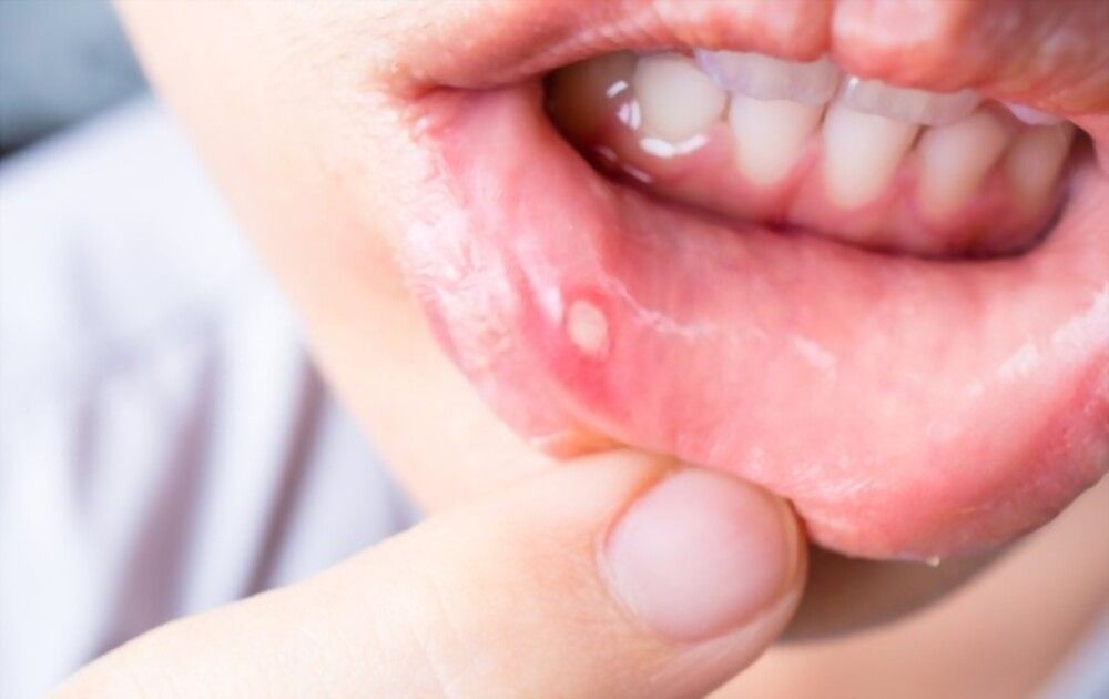
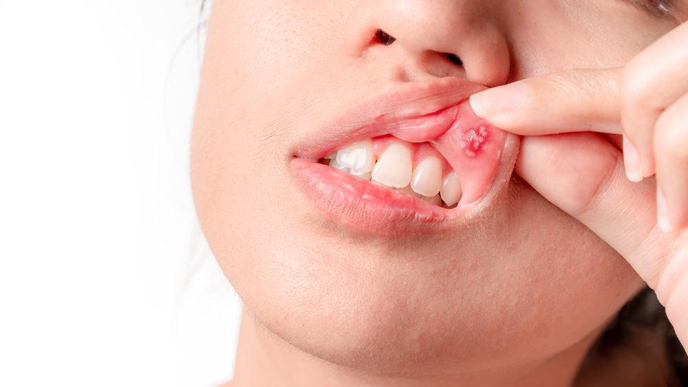
Mouth ulcers, also known as canker sores are normally small, painful lesions that develop in your mouth or at the base of your gums. They can make eating, drinking, and talking uncomfortable.
Women, adolescents, and people with a family history of mouth ulcers are at higher risk for developing mouth ulcers.
If you get a canker sore that is large or extremely painful, or if it lasts for a long time without healing, you should seek the advice of a doctor.
Mouth Ulcers do not need treatment as they go away with time. But if the ulcers are large and extremely painful please visit your dentist for mouth ulcer treatment options:
· Rinse with salt water and baking soda
· Apply pain relief ointments like Orajel or Anbesol
· Apply ice to canker sores or use cold water
· Placing old tea bags on the sores
· Drink cool chamomile tea
· Milk of magnesia can also be a good option and is advised to place on Mouth Ulcers
Mouth ulcers need to be treated at their early stages by experts in oral medicine and radiology. For the best treatment, you can visit Dental World Super-SpecialityClinic, one of the best Oral Medicine and Radiology clinic in Kolkata.
Mouth ulcers can be easily treated at home by rinsing the mouth regularly with warm salt water, drinking plenty of fluids, avoiding spicy food and applying an antiseptic gel to the ulcers. Usually, mouth ulcers heal within a week. In case they don’t, consider visiting your nearest dentistin Kolkata.
Mouth ulcers usually self heal within one to two weeks. However, serious and painful ulcers can take up to 6 weeks to heal. If you are prone to mouth ulcers, visit the best Oral Medicine and Radiologist in Kolkatafor immediate relief from painful canker sores.)
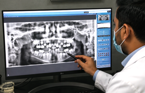
X-rays use radiation to take pictures of bones and other parts inside the body. An OPGxray is a panoramic X-ray of the upper and lower jaws, including the teeth. The OPG machine is specifically designed to rotate around the patient’s head during the scan. An OPG will take approximately 20 seconds.
An OPG can be used to look for
Your doctor, dentist or dental specialist knows the risks of having an OPG and will consider the risks before recommending you have this type of X-ray.
Possible risks are
Bring your referral letter or request form and all OPG X-rays taken within the last 2 years with you. Leave the X-rays with the medical imaging staff as the doctor may need to look at them. The staff member will tell you when these are ready to be picked up.
Leave all jewellery and valuables at home.
There is no special preparation for an OPG.
You may be asked to remove any metal objects.
Important to tell your doctor before the OPG
If you are or may be pregnant.
Medical imaging staff will ask you to sit on a chair or stand for the OPG. It is important that you tell the staff if you have difficulty sitting or standing unassisted. The radiographer may place a protective shield over the parts of your body not being X-rayed, or you may be asked to wear a protective apron.
When you are ready, the radiographer will go behind a screen to start the OPG machine. They will ask you to be still during the OPG. When your OPG is finished you will be asked to wait while the radiographer checks the pictures. The procedure usually takes about 5 minutes including time taken to get ready.
The amount of time it takes for you to get your results will differ depending on where you get your scans done. The medical imaging doctor will look at the images and issue a report. The images may be on films on a CD or on a computer. Ask whether you should wait to take the images and report with you, or whether they will be sent to your doctor or dentist. You will need to make an appointment to discuss the images and report with your doctor or dentist.
You will be able to go soon after the OPG is finished and can continue with normal activities.
The amount of time it takes for you to get your results will differ depending on whereyou get your scans done. The department of Oral Medicine and Radiology will look at the images and issue a report. The images may be on films on a CD or on a computer. Ask whether you should wait to take the images and report with you, or whether they will be sent to your doctor or dentist. You will need to make an appointment with an Oral Medicine and Radiology Clinic in Kolkata to discuss the image & report.
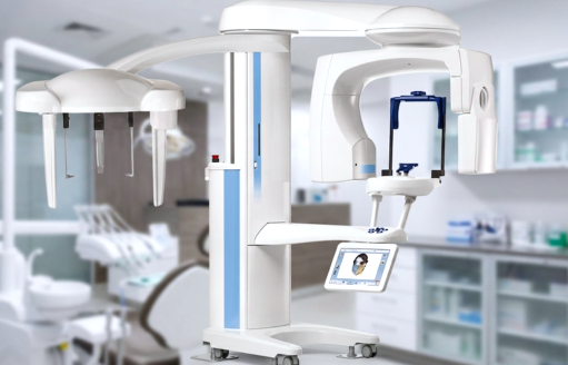
Cone beam computed tomography is a medical imaging technique consisting of X-ray computed tomography where the X-rays are divergent, forming a cone. CBCT has become increasingly important in treatment planning and diagnosis in implant dentistry, ENT, orthopedics, and interventional radiology, among other things.
Cone-beam computed tomography systems (CBCT) are a variation of traditional computed tomography (CT) systems. The CBCT systems used by dental professionals rotate around the patient, capturing data using a cone-shaped X-ray beam.
Dental CBCT systems have been sold in the United States since the early 2000s and are increasingly used by radiologists and dental professionals for various clinical applications including dental implant planning, visualization of abnormal teeth, evaluation of the jaws and face, cleft palate assessment, diagnosis of dental caries (cavities), endodontic (root canal) diagnosis, and diagnosis of dental trauma.
about 20-40 seconds
During a CBCT scan, the imaging machine rotates entirely around the patient's head. In less than a minute, about 150-200 images are captured from a variety of angles and compiled into a single 3D image. CBCT scans are quick and in most cases, a full mouth scan only takes about 20-40 seconds.
No, the CBCT test is not at all painful. In a sense, you can liken it to the experience of getting photographed, which causes no pain or discomfort.
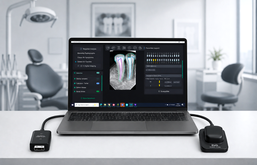
RVG stands for RadioVisioGraphy. This technology is used as an alternative to X-ray radiography in dentistry. RVG Sensors are developed keeping their durability and image quality in mind. The hermetically sealed sensor housing makes this sensor waterproof allowing for versatile applications.
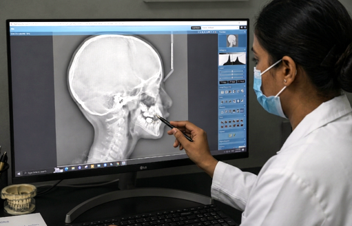
A Lat Ceph is a lateral or side view x-ray of the face, which demonstrates the bones and facial contours in profile on a single film. Lat CEPH x-rays are usually used in the diagnosis and treatment of orthodontic problems.
1st Floor, 204, Acharya Jagdish Chandra Bose Rd, Mullick Bazar, Park Street area, Kolkata, West Bengal 700017
Ph : +91 9073336669
Ground Floor, Salt Lake Merlin matrix, 10, DN Block, Sector V, Bidhannagar, Kolkata, West Bengal 700091
Ph : +91 9073336669